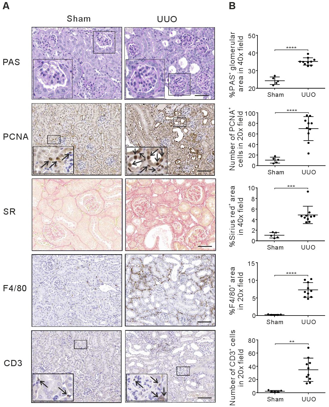
Fig. 8. Markers of fibrosis and inflammation in New Zealand obese (NZO) mice. (A) Periodic acid Schiff (PAS) stain: marker of glomerular damage (40x). Scale bar: 50 μm. Proliferating cell nuclear antigen (PCNA) antibody stain: marker of cell regeneration (20x. Scale bar: 100 μm; arrows show positive cells). Sirius red (SR) stain: marker of interstitial fibrosis (40x. Scale bar: 50 μm). F4/80 antibody stain: macrophage marker (20x. Scale bar: 100 μm) and CD3 antibody stain: T-cell marker (20x. Scale bar: 100 μm; arrows show positive cells) in sham-operated group and UUO-operated groups. (B) All quantification data are means ± SD. Data were determined in n=5 for sham-operated kidneys, n=10 for UUO-operated kidneys. **p<0.01, ***p<0.001 and ****p<0.0001.